
Lung Structure BioNinja
1/6 Synonyms: none The majority of biochemical reactions that occur within the body are dependent on energy. In order to create these energy moieties, the cellular organelles utilize oxygen to drive these chemical reactions. The human lungs are a pair of spongy organs within the thoracic cavity that facilitate gaseous exchange.
Lungs clipart labeled, Lungs labeled Transparent FREE for download on WebStockReview 2023
Health tips Overview The lungs are the center of the respiratory (breathing) system. Every cell of the body needs oxygen to stay alive and healthy. Your body also needs to get rid of carbon.

Lung Diagrams Diagram Link
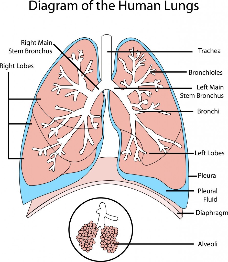
The current article provides a labeled diagram of the human lungs as well as a description of the parts and their functions. The blood-air barrier in the alveoli is 50 times thinner than a sheet of trace paper. This thinness facilitates faster oxygenation.
Diagram of lungs
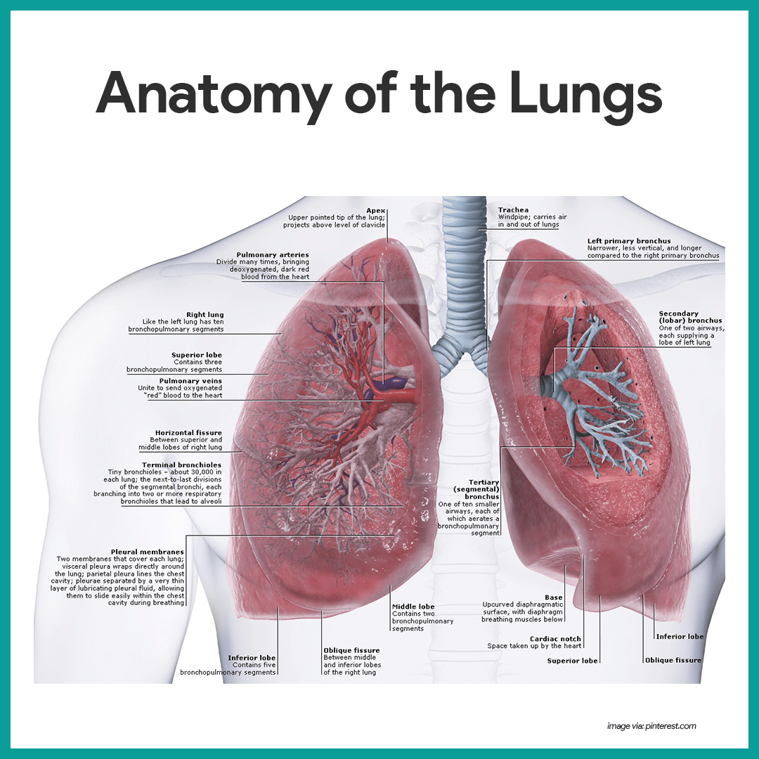
The diaphragm is the flat, dome-shaped muscle located at the base of the lungs and thoracic cavity. The lungs are enclosed by the pleurae, which are attached to the mediastinum. The right lung is shorter and wider than the left lung, and the left lung occupies a smaller volume than the right.
Lung Diagram With Labels General Wiring Diagram
The lungs are a major organ that is part of the respiratory system, taking in fresh air and getting rid of old, stale air. This mechanism of breathing also helps to allow you to talk. By taking in fresh air, the lungs are able to help oxygenate blood to be carried around your body. This is done by inhaling the air and bringing it in toward the.
Lung Diagram Labeled EdrawMax Template
Recommended video: Respiratory system [23:23] Main structures of the respiratory system. Respiratory system (Systema respiratorum) The respiratory system, also called the pulmonary system, consists of several organs that function as a whole to oxygenate the body through the process of respiration (breathing).
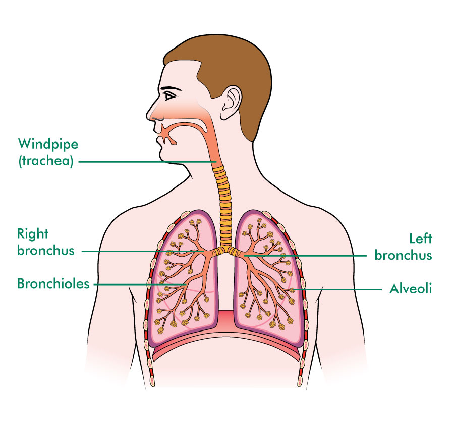
The lungs Lung cancer Macmillan Cancer Support
Anatomy, Thorax, Lungs - StatPearls - NCBI Bookshelf The hilum (root) is a depressed surface at the center of the medial surface of the lung and lies anteriorly to fifth through seventh thoracic vertebrae. It is the point at which various structures enter and exit the lung.
Respiratory System Anatomy and Physiology Nurseslabs
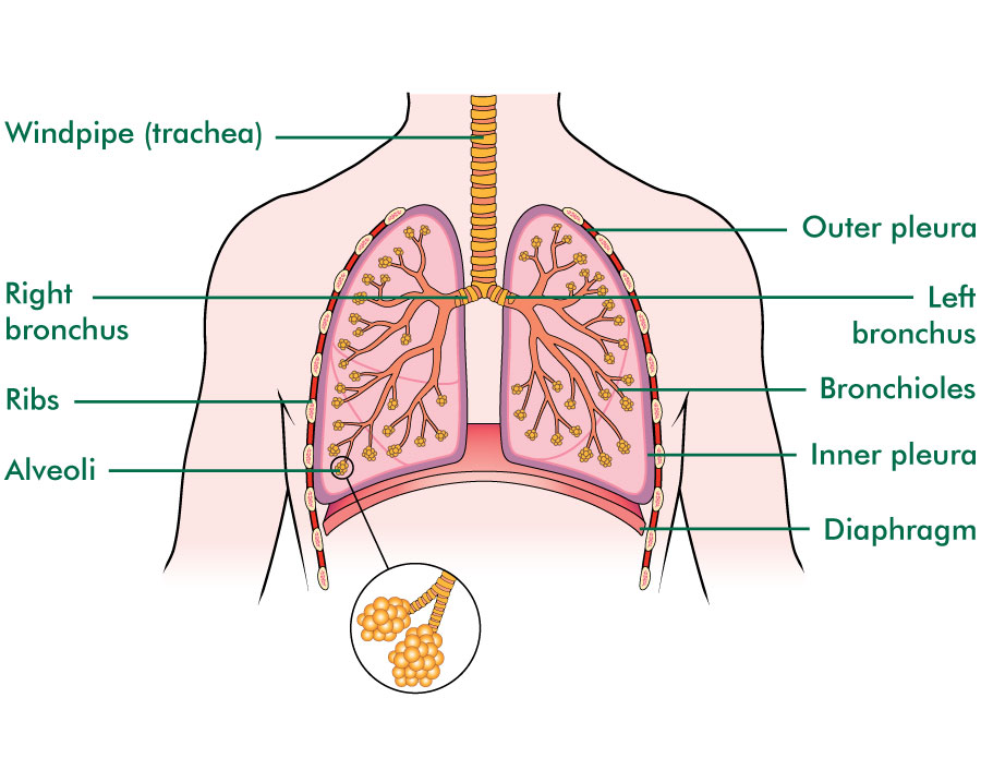
Respiratory system diagram The respiratory system How we breathe Respiratory conditions Summary The respiratory system allows air to reach the lungs, from which oxygen enters the blood.

Lungs and Heart (Posterior View, labelled), illustration Stock Image C043/4864 Science
Total Lung Capacity (TLC) and Lung Compliance. TLC refers to the maximum volume of air the lungs of an adult person can hold. It is the sum of the air released by the lung after a maximum exhalation (vital capacity or VC) and the volume of air left behind within the lungs after a deepest exhalation (residual volume or RV) [46].The TLC of human lungs is 6 liters [47].

Labeled diagram of the lungs/respiratory system.
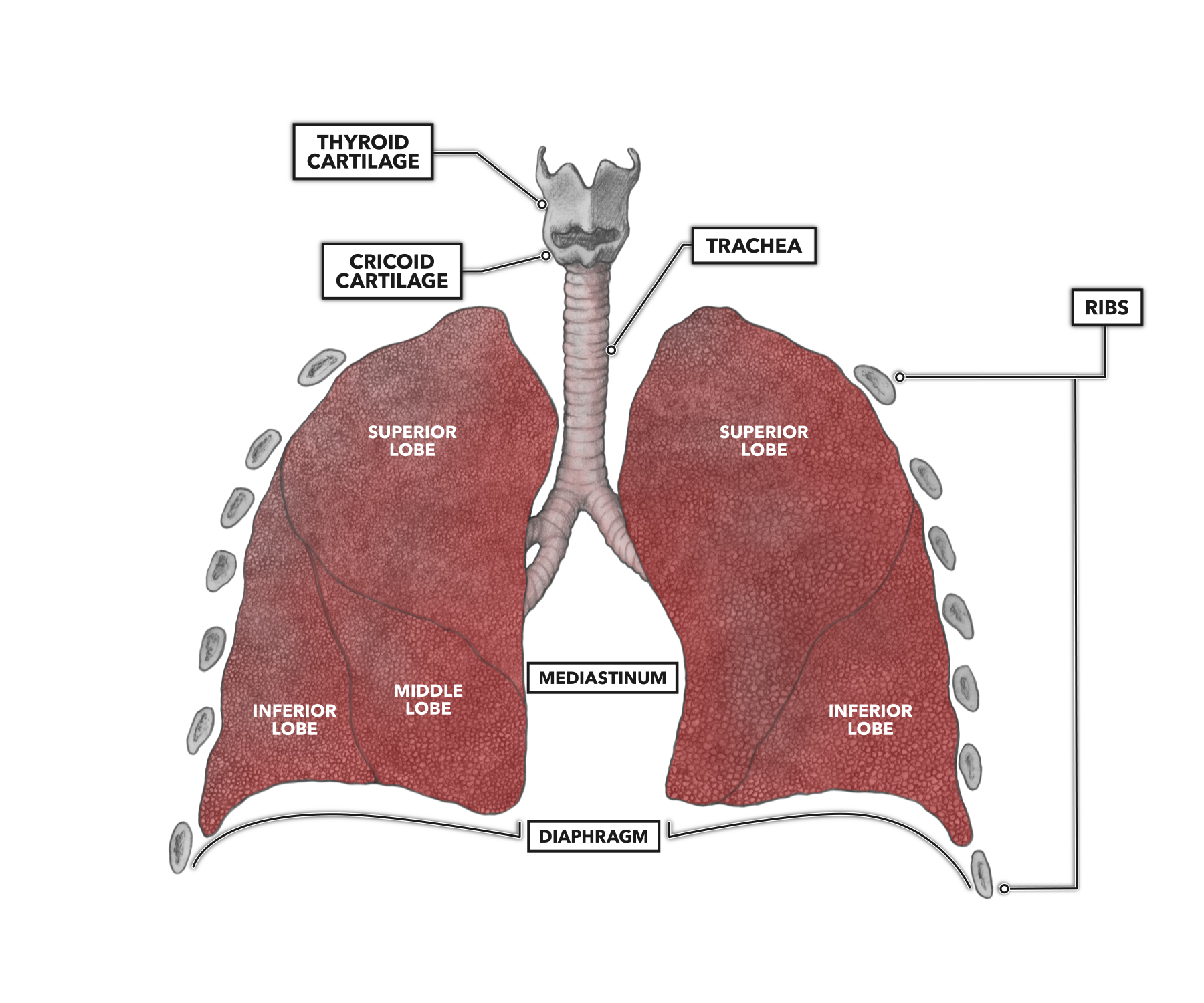
Lungs are a pair of respiratory organs situated in a thoracic cavity. Right and left lung are separated by the mediastinum. Texture -- Spongy Color - Young - brown Adults -- mottled black due to deposition of carbon particles Weight- Right lung - 600 gms Left lung - 550 gms THORACIC CAVITY SHAPE - Conical Apex (apex pulmonis) Base
CrossFit Anatomy of the Lungs
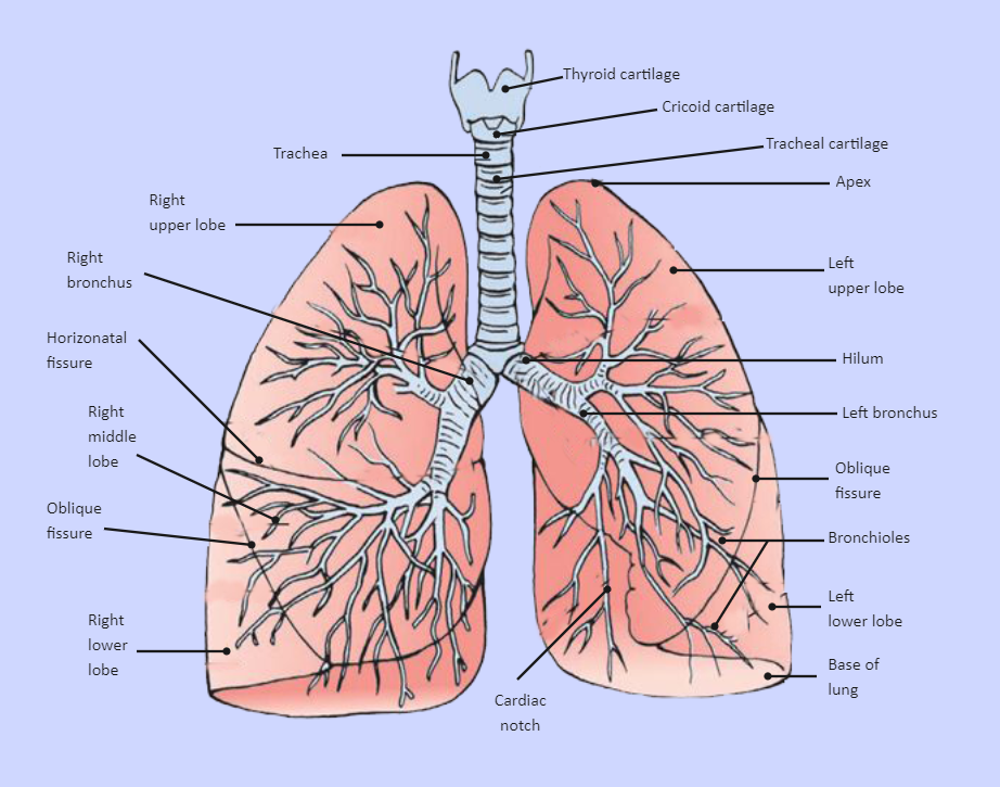
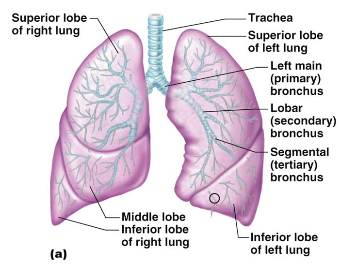
Lungs-lobes: diagrams detailing the various lung lobes (lower lobe, middle-right lobe, left lingula) separated by the pulmonary fissures. Hilum of lung - Root of lung - Superior lobe; Upper lobe - Lingula of left lung - Middle lobe of right lung - Inferior lobe; Lower lobe
A healthy lung has a pinkish appearance, and if you could see it outside the body, it would look
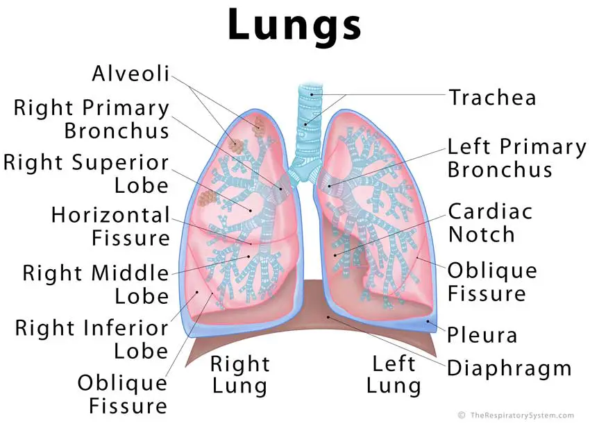
Pleura of the Lungs. Each lung is enclosed within a cavity that is surrounded by the pleura. The pleura (plural = pleurae) is a serous membrane that surrounds the lung. The right and left pleurae, which enclose the right and left lungs, respectively, are separated by the mediastinum. The pleurae consist of two layers.
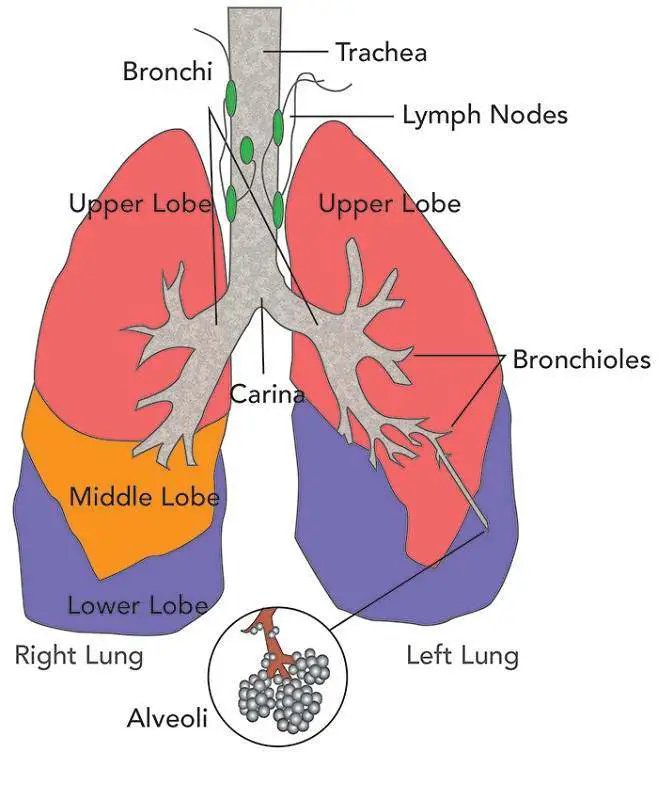
Diagram of the lungs
The respiratory system aids the body in the exchange of gases between the air and blood, and between the blood and the body's billions of cells. It includes air passages, pulmonary vessels, the.

right lung model A&P.4.Heart.Lung Pinterest Lungs and Nursing students
The reduction of surface tension means a reduction of work of breathing. The surfactant is not static but continually being turned over and removed. The removal is by pinocytotic vesicles of type II pneumocytes, macrophages, and type I pneumocytes.

2 Embryology, anatomy, and physiology of the lung Thoracic Key
Revisions: 49 format_list_bulleted Contents add The lungs are the organs of respiration. They are located in the thorax, either side of the mediastinum. The function of the lungs is to oxygenate blood. They achieve this by bringing inspired air into close contact with oxygen-poor blood in the pulmonary capillaries.
CLASS BLOG BIO 202 Respiratory System KEY
Labeled diagram of the lungs/respiratory system. View Original Image at Full Size Labeled diagram of the lungs/respiratory system. Image 37789 is a 1125 by 1408 pixel WebP Uploaded: Jan10 14 Last Modified: 2014-01-10 12:15:34 Permanent URL: https://serc.carleton.edu/download/images/37789/labeled_diagram_lungsrespirato.v2.webp